| home | virtual field trips | regional geology | images | e-learning | links |
Images on the scanning electron microscope (SEM) are generated as a fine electron beam scans across a sample. This interaction results in release of energy as electrons, X-rays, and light, which are recorded by various detectors inside the microscope. The signals from the detectors are then acquired as digital images; formerly, they were recorded on photographic film. Two main types of image are of interest to geologists:
Secondary electron images (SEI) are built up from low energy secondary electrons released from the sample by the electron beam. Such images emphasize the topography of the sample. Samples are typically loose grains, small rock samples or fossils, mounted on an aluminium stub and given a thin gold coat.
Back-scattered electron images (BEI) are built up from high energy electrons from the electron beam, which are back-scattered from the sample. The specimen is a thin section or block, polished flat, with a thin carbon coat. BEIs emphasize compositional differences across the sample.
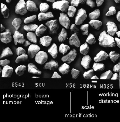 |
In this secondary electron image of sand grains, we see a topographic representation of the sand grains, seen as if illuminated from the top left. The data at the bottom of the photograph are explained. The most important are:
Use the scale rather than the magnification. Magnification is only correct if you are viewing the image at its original size, and this is often not the case. |
![]()
![]()
This page is maintained by Roger Suthren. Last updated 22 November, 2021 12:04 PM